Diagnostic Imaging
This page is the place to find clear and reliable information about the imaging techniques used by our veterinarians. From conventional radiographs to advanced CT and MRI scans, you will discover how these examinations work and the role they play in diagnosing disease.
Our veterinarians share their expertise in clear, well-structured articles, grouped by theme. This makes it easy to navigate and quickly find the examinations that are relevant for your pet.


Radiography
Radiography is one of the most commonly used techniques to obtain an initial assessment of the skeleton, thorax, or abdomen. Here you can learn more about the applications and variants of this examination.
Standard radiographs provide rapid insight into bony structures and organ positioning. They are frequently used to detect fractures, pulmonary abnormalities, and changes within the thoracic or abdominal cavity. The examination is widely applicable and relatively quick to perform. Radiography often represents the first step in the diagnostic process.
Dental radiography is essential for assessing dental and jaw disorders. The images reveal structures that are not visible during a clinical examination, such as tooth roots and jawbone. This allows detection of underlying infections, resorptive lesions, or fractures. The examination supports accurate diagnosis and targeted dental treatment.
Fluoroscopy is an imaging technique that allows real-time visualization of movement. It is used, among other indications, to investigate swallowing disorders and respiratory problems. This enables detection of functional abnormalities that cannot be assessed with standard radiographs. The technique provides valuable information for diagnosis and further treatment.
Contrast studies use contrast agents to enhance visualization of organs and structures during imaging. They are commonly applied in examinations of the gastrointestinal tract, urinary system, or blood vessels. This allows clearer identification of strictures, leaks, or displacements. Contrast imaging is a valuable adjunct to standard radiography.
Hip, elbow, and shoulder dysplasia are detected using standardized radiographic examinations. These screenings allow objective assessment of joint abnormalities. They play an important role in breeding decisions and early detection of orthopedic problems. In addition, the results can help guide further follow-up or treatment.

Ultrasound
Ultrasound uses sound waves to create detailed images of organs, muscles, and blood vessels. In this section, you can read how this technique is applied in veterinary medicine.
A standard ultrasound examination provides insight into the organs of the abdomen and thorax. Abdominal and thoracic ultrasound allow assessment of organ structure, size, and blood supply. The examination is non-invasive and can be performed in real time. Ultrasound is an important tool for establishing an accurate diagnosis.
Musculoskeletal ultrasound is used to evaluate muscles, tendons, and joints. The examination allows visualization of injuries, inflammation, and structural changes. Movement can be assessed during the examination, providing additional functional information. This technique supports targeted diagnosis and treatment planning.
Doppler ultrasound is used to assess blood flow within blood vessels and organs. The technique provides information on the direction and velocity of blood flow. It is indicated when vascular problems or circulatory disorders are suspected. Doppler ultrasound is an important complement to standard ultrasound examination.
Ultrasound is a safe and reliable method for confirming pregnancy in various animal species. The examination allows assessment of the presence and development of embryos. It can also provide information on viability and progression of pregnancy. Pregnancy diagnosis supports appropriate monitoring throughout gestation.

Video Endoscopy
Endoscopy allows the internal examination of the airways, gastrointestinal tract, or other organs. It is a valuable technique for both diagnosis and treatment.
Diagnostic endoscopy makes it possible to directly visualize internal structures such as the nasal cavity, airways, esophagus, stomach, or bladder. The examination provides detailed views of mucosal surfaces and abnormalities. If necessary, samples can be collected during the procedure. Endoscopy delivers valuable information using a minimally invasive approach.
In addition to diagnostic purposes, endoscopy can also be used therapeutically. Techniques such as laparoscopy and thoracoscopy enable minimally invasive procedures. This often results in less tissue damage and faster recovery. The method is applied in both surgical treatments and targeted interventions.
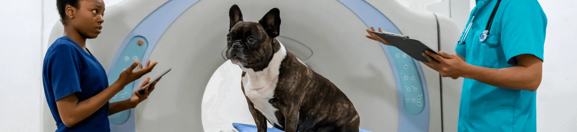
CT and MRI Scans
A CT scan creates cross-sectional images of the body and provides highly detailed views of bones, organs, and soft tissues. An MRI scan uses magnetic fields to produce very detailed images, particularly of the brain, spinal cord, and joints.
An MRI scan uses magnetic fields to generate highly detailed images, especially of the brain, spinal cord, and joints. This technique is frequently used in the evaluation of neurological and orthopedic conditions.
A CT scan produces detailed cross-sectional images of the body. The examination is particularly well suited for visualizing bony structures, organs, and soft tissues. CT is often used in cases of trauma, tumors, and complex anatomical abnormalities. The images provide rapid and accurate diagnostic information.